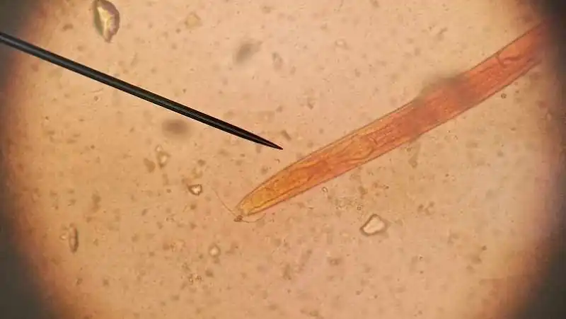
With the gradual advancement of modern intensive farming, swine diseases remain complex. Swine strongyloidiasis is a parasitic disease caused by Strongyloides ransomi, which poses a significant threat to piglets. It can occur year-round, with a higher incidence in the hot and humid summer. The parasites mainly infect the host through two routes: oral ingestion and skin penetration. Infected pigs primarily exhibit poor growth and development, persistent diarrhea, and respiratory symptoms, which can lead to death in severe cases.

I. Epidemiology
There are two main routes of infection in swine herds: oral infection and skin penetration. Once filariform larvae in the environment contaminate feed and drinking water, healthy pigs can ingest them, leading to infection in the intestines. Suckling piglets and piglets in the early nursery stage are particularly susceptible as they are prone to consuming feed on the ground, resulting in oral infection. After ingestion, the filariform larvae can penetrate the gastric mucosa and enter the bloodstream, traveling to the lungs via blood circulation. From the lungs, they migrate up the trachea to the pharynx and are then swallowed, reaching the small intestine where they develop into adult worms.
Due to their delicate skin, piglets are also susceptible to skin penetration by environmental larvae. The larvae actively penetrate the skin, burrowing into the subcutaneous tissue and muscles. From there, they travel through blood and lymphatic vessels to the lungs, then up the trachea with mucus to the pharynx, are swallowed, and reach the small intestine, where they develop into parthenogenetic female worms. The period from infection to egg laying by the parasitic female worms is approximately 10-12 days, and these females can survive in the host for 6-9 months.
Strongyloidiasis primarily affects piglets. Eggs can be detected in the feces of piglets as early as 5-8 days after birth, with the infection being most severe around 1 month of age and gradually decreasing after 2-3 months. Diseased pigs and subclinical carriers are the main sources of infection. The disease can occur year-round, with the highest incidence in the hot and humid summer. Pig farms with poor hygiene are most susceptible. Without treatment, the mortality rate can reach 50%.
II. Clinical Symptoms
Strongyloides ransomi mainly affects piglets. When the larvae invade and infect piglets through the skin, the skin experiences a certain degree of irritation, generally manifesting as rashes and dermatitis. When the larvae invade and parasitize the lungs, they can obstruct the small bronchioles or bronchioles near the parasitic sites, causing pinpoint hemorrhages. This subsequently leads to inflammation in these areas, clinically presenting as bronchopneumonia and pleuritis. If the larvae inadvertently enter the heart through the pulmonary vessels during their migration, or enter the brain tissue from the heart vessels, it can lead to acute death in piglets.
When a large number of worms parasitize the small intestine, it can cause parasitic enteritis, characterized by thickened, desquamated, and hemorrhagic mucous membranes. Insufficient secretion of digestive enzymes and inadequate digestion and absorption of feed lead to nutritional deficiencies. The symptoms in affected pigs include digestive disorders, abdominal pain, diarrhea with blood and sloughed intestinal mucosa in the feces.
III. Differential Diagnosis
- Fecal examination using the saturated salt flotation method: Fresh feces must be used (not exceeding 6 hours in summer). Oval-shaped eggs containing coiled larvae will be visible.
- Post-mortem examination: Due to the small size of the worms and their location deep within the small intestinal mucosa, it is necessary to scrape the mucosa with a scalpel and carefully examine it in clear water to detect the parasites.
IV. Treatment and Prevention Measures
Treatment:
- For pigs with rashes and dermatitis, apply a solution of iodine tincture (1:50) to the inflamed areas. Combine this with intramuscular injections of sensitive antibiotics such as cephalosporins and enrofloxacin. Generally, 3-5 days of treatment can effectively treat and control the condition.
- For pigs with severe diarrhea, fluid replacement therapy is essential, followed by antibiotics such as a combination of “Wuti Tongzhi” (五体统治) at 2-3 kg/ton of feed + chlortetracycline at 2 kg/ton of feed for preventing or treating secondary infections. For piglets with fever, antipyretics should be administered promptly. For pigs with severe abdominal breathing, administer kanamycin at 40,000 IU/kg body weight via intramuscular injection. Recovering pigs should be supplemented with “Jianeng Subu” (加能速补) for conditioning.
Prevention:
Since the eggs and larvae have very low resistance to dryness, it is crucial to maintain dry pens and promptly remove pig manure. Simultaneously, regular preventive deworming of the pig herd should be carried out. “Chonglihei” (虫力黑) can be used at a rate of 1 kg/ton of feed for a “four plus one” deworming regimen: sows are dewormed four times a year, and meat pigs are dewormed once around 60 days of age (with another deworming around 120 days of age for larger pigs).
